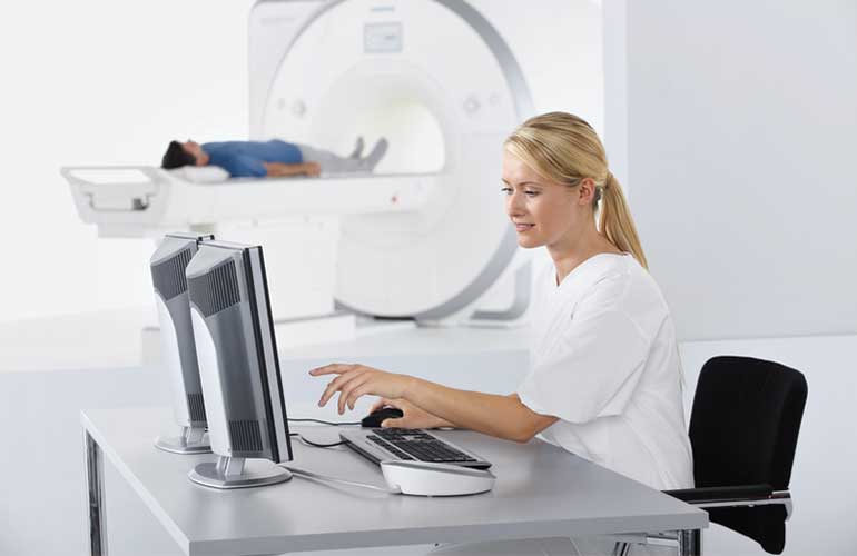
Magnetic resonance imaging (MRI)
MRI is a diagnostic examination in which pulses of radio waves in a magnetic field stimulate different tissues of the body to emit weak radio signals, which are picked up by sensitive antennas (coils). A computer processes these signals and converts them into images. There is no X-ray (ionising) radiation during an MRI examination. To date, no harmful effects of the examination are known.
Book an appointmentDo you need to prepare for the examination?
General guidelines apply to MRI examinations, and every patient should be familiar with them before having one. MRI of certain organs also requires additional specific preparation (described under the types of MRI examinations).
The magnetic field of the MRI machine is strong and always active! Certain metal implants, devices or objects near the MRI machine can therefore be life-threatening for you.
If you have metal foreign bodies or other implants, you must obtain a certificate or your attending physician's opinion on the compatibility of the implant or device with an MRI examination. For safety reasons, we do not perform MRI examinations at our facility on people with an implanted pacemaker or defibrillator.
Before entering the examination room, you must remove ALL removable metal objects, including hearing aids, dentures, keys, mobile phones and other electronic devices, glasses, hairpins, headwear, jewellery, piercings, watches, belts, safety pins, paper clips, bank and credit cards, parking tickets with a magnetic strip, coins, pens, pocket knives, nail clippers, lighters, clothing with metal clasps, press studs or zips, and other such items.
Wear clothing without metal buttons or fasteners.
How is the examination performed?
During the procedure, you lie in the opening of the MRI machine, which is shaped like a tunnel. Some people may feel a sense of confinement because of this.
You will hear loud knocking noises at intervals. You will be given headphones or earplugs to reduce the noise.
It is important that you remain completely still during the examination, as even the slightest movement of the body part being examined can significantly reduce its diagnostic value. If pain makes it difficult for you to lie still on your back, you may take a painkiller before the procedure.
In certain cases, administering a contrast agent significantly improves the informative value of the examination. On the radiologist's instructions, the patient receives it as an intravenous injection; only in MR arthrography is the contrast agent injected into the joint being examined.
The examination takes from 15 minutes to one hour, depending on the part of the body being examined and whether a contrast agent is used.
An MRI examination is painless for patients.
Are complications possible with the contrast agent?
When the contrast agent is injected, you may feel a sensation of cold at the injection site; this is expected and is not a complication.
A small bruise may appear at the injection site.
In very rare cases, an allergic reaction to the contrast agent may develop, most likely within the first half hour. It manifests as nausea, itching, a rash or fluctuations in blood pressure. ALERT US IMMEDIATELY if you notice any of these signs!
Severe allergic complications, such as difficulty breathing, convulsions, a drop in blood pressure and heart rhythm disturbances, are very rare. The probability of their occurring is 1 in 10,000. The risk is negligible compared with the benefit of the examination.
In severe kidney impairment, the contrast agent may be deposited in other tissues of the body, with consequent harmful effects. If you have impaired kidney function, tell us before the contrast agent is administered!
It is very important that you tell us before your visit about any conditions that may affect the course of the examination, namely:
- a known allergy to contrast agents or a previous severe allergic reaction,
- fear of enclosed spaces (claustrophobia),
- kidney disease,
- pregnancy.
If you have any doubts or questions about this, consult our staff BEFORE YOU ENTER the examination room!
MRI questionnaires
Before an MRI examination, you need to complete the general MRI questionnaire and, if you are having a breast MRI, the breast MRI questionnaire as well. To speed up your visit, please bring the appropriate completed questionnaire with you.
Speed up your visit. Complete the questionnaire, available here, before coming to the clinic.
General MRI questionnaire Breast MRI questionnaireTypes of examinations
We offer a wide range of MRI examinations. The general MRI guidelines apply to every examination, while individual examinations also require additional specific preparation, described below.
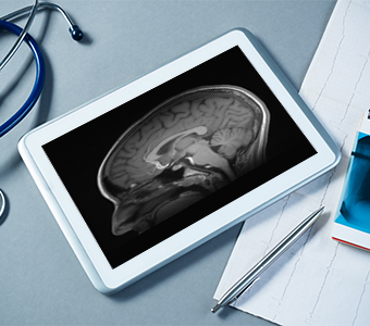
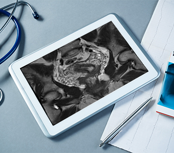
MRI of the head and neck
Do you need to prepare for the examination?
If you are scheduled for an MRI of the head with a contrast agent (for example, to characterise tumours or as part of the diagnosis of multiple sclerosis), do not eat for at least 6 hours before the examination.
In other cases, no special preparation is needed for an MRI of the head; only the general MRI guidelines apply.
How is the examination performed?
The radiographer will position you appropriately on the examination table and fit a suitable coil. You will also be given headphones to protect your hearing, as loud noises are heard during the examination. You will then be moved into the MRI machine for the duration of the scan. You will also be given a call bell, which you can press if you experience any problems during the procedure to contact the radiographer carrying it out.
It is especially important that you keep completely still during the examination itself, as movement degrades the quality of the images!
The examination takes about 30 minutes. If a contrast agent also needs to be administered, this adds a further 15 minutes.
Is a contrast agent required?
A contrast agent is not routinely used in these examinations, except in special cases, such as the characterisation of known tumours, vascular malformations, the diagnosis of multiple sclerosis, etc.
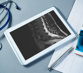
MRI of the spine
Do you need to prepare for the examination?
If you have been referred for an MRI of the spine with a contrast agent (for example, to characterise tumours, as part of the diagnosis of multiple sclerosis, after previous surgery, etc.), do not eat for at least 6 hours before the examination.
Otherwise, no special preparation is needed; only the general MRI guidelines apply.
How is the examination performed?
The radiographer will position you appropriately on the examination table and fit a suitable coil for the part of the spine being examined. You will also be given headphones to protect your hearing, as loud noises are heard during the examination. You will then be moved into the MRI machine for the duration of the scan. You will also be given a call bell, which you can press if you experience any problems during the procedure to contact the radiographer carrying it out.
It is especially important that you keep completely still during the examination itself, as movement degrades the quality of the images!
The examination takes around 15–30 minutes. If a contrast agent also needs to be administered, this adds a further 15 minutes.
Is a contrast agent required?
A contrast agent is not routinely used in these examinations, except in special cases, such as the characterisation of known tumours, multiple sclerosis, or after previous surgery.
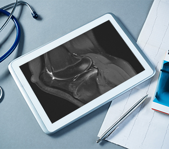
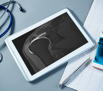
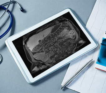
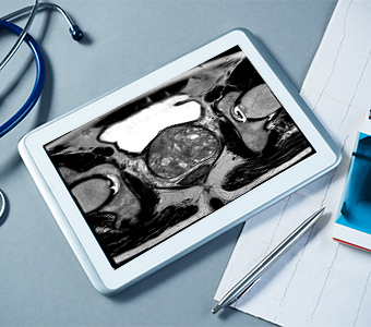
MRI of the joints and limbs
Do you need to prepare for the examination?
No special preparation is needed for examinations of the joints and limbs; only the general MRI guidelines apply.
How is the examination performed?
The radiographer will position you appropriately on the examination table and fit a coil for the part of the body being examined. You will also be given headphones to protect your hearing, as loud noises are heard during the examination. You will then be moved into the MRI machine for the duration of the scan. You will also be given a call bell, which you can press if you experience any problems during the procedure to contact the radiographer carrying it out.
It is especially important that you keep completely still during the examination itself, as movement degrades the quality of the images!
The examination takes around 15–30 minutes. If a contrast agent also needs to be administered, this adds a further 15 minutes.
Is a contrast agent required?
A contrast agent is not routinely used in these examinations, except in special cases, such as the characterisation of known tumours or in septic inflammation.
MR arthrography
What is MR arthrography?
MR arthrography is an examination in which a contrast agent is injected into the joint being examined. This improves the contrast between the joint fluid and the structures inside the joint. It also increases the volume of fluid in the joint, which further opens up small injuries to the intra-articular structures.
MR arthrography is the most sensitive examination for injuries to structures inside the joint (the labrum, rotator cuff tendons, cartilage).
Do you need to prepare for the examination?
No special preparation is needed for MR arthrography of the joints; only the general MRI guidelines apply.
How is the examination performed?
MR arthrography takes place in two stages.
The first stage is carried out in the ultrasound room. The doctor will position you appropriately on the examination table, clean the area of the planned puncture with a disinfectant and cover it with sterile drapes. Under ultrasound guidance, they will then puncture the joint being examined with a thin needle and inject the contrast agent into it. The pain of the procedure is comparable to that of having blood drawn from a vein. The first stage takes about 5–10 minutes.
In the second stage, imaging is performed in the MRI machine; for the patient, this is the same as a standard MRI examination. The radiographer will position you appropriately on the examination table and fit a coil for the part of the body being examined. You will also be given headphones to protect your hearing, as loud noises are heard during the examination. You will then be moved into the MRI machine for the duration of the scan. You will also be given a call bell, which you can press if you experience any problems during the procedure to contact the radiographer carrying it out.
It is especially important that you keep completely still during the examination itself, as movement degrades the quality of the images!
The second stage takes about 15–30 minutes.
Is a contrast agent required?
In MR arthrography, a solution is injected into the joint being examined in which the gadolinium contrast agent is diluted 1,000-fold in saline.
Are complications possible after the examination?
A small bruise may form at the puncture site.
After the examination, mild pain or a feeling of discomfort may occur, but these are usually not pronounced and subside within a few days at most.
An allergic reaction is very rare, as extremely low concentrations of contrast agent are used in MR arthrography.
Joint infection is also very rare, occurring in fewer than 1 in 10,000 cases.
If you experience severe or unexpected problems, let us know immediately!
MRI of the upper abdomen
An MRI of the upper abdomen covers the liver, gallbladder, bile ducts, pancreas, spleen, adrenal glands and kidneys.
Do you need to prepare for the examination?
You must not eat for at least 6 hours before the examination. If you have been referred for an MRI of the gallbladder, bile ducts or pancreas, do not drink anything for the last 4 hours before the examination. Otherwise, you may drink only small amounts of water.
In all other respects, the general MRI guidelines apply.
How is the examination performed?
The radiographer will position you appropriately on the examination table and fit a coil for the part of the body being examined. You will also be given headphones to protect your hearing, as loud noises are heard during the examination. You will then be moved into the MRI machine for the duration of the scan. You will also be given a call bell, which you can press if you experience any problems during the procedure to contact the radiographer carrying it out.
It is especially important that you keep completely still during the examination itself, as movement degrades the quality of the images!
The examination takes up to 45 minutes. If a contrast agent also needs to be administered, this adds a further 15 minutes.
Is a contrast agent required?
In these examinations, a contrast agent is used as needed, for example for more precise characterisation of tumours, in inflammation, etc. The decision is made by the radiologist based on your clinical information and the findings during the examination itself.
MR enterography
What is MR enterography?
MR enterography is a non-invasive radiological method used to image the small bowel. It can reveal changes associated with chronic inflammatory bowel disease, tumour growths, post-operative scarring, etc.
Do you need to prepare for the examination?
Do not eat for 8 hours before the examination. Do not drink any liquids for the last 4 hours.
At our facility, you will receive 1.5 litres of liquid contrast agent 45–60 minutes before the examination, which you will drink slowly until the scan begins. During this time, we will also insert an intravenous line, through which you will receive a medication to relax the muscles of the bowel wall and a contrast agent during the examination.
In all other respects, the general MRI guidelines apply.
How is the examination performed?
The radiographer will then position you appropriately on the examination table and fit a coil. You will also be given headphones to protect your hearing, as loud noises are heard during the examination. You will then be moved into the MRI machine for the duration of the scan. You will also be given a call bell, which you can press if you experience any problems during the procedure to contact the radiographer carrying it out.
It is especially important that you keep completely still during the examination itself, as movement degrades the quality of the images!
Including preparation, the examination takes about 2 hours.
Is a contrast agent required?
Yes, an intravenous contrast agent is used in MR enterography.
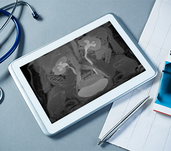
MR urography
What is MR urography?
MR urography is a non-invasive diagnostic examination used to image the urinary tract. It can identify congenital anatomical anomalies of the urinary tract, any narrowing of the ureters, tumours and their extent, etc.
Do you need to prepare for the examination?
You must not eat for at least 6 hours before the examination. You may drink moderate amounts of liquid.
Half an hour before the examination, we will insert an intravenous line, through which you will receive saline solution.
In all other respects, the general MRI guidelines apply.
How is the examination performed?
The radiographer will position you appropriately on the examination table and fit a coil for the part of the body being examined. You will also be given headphones to protect your hearing, as loud noises are heard during the examination. You will then be moved into the MRI machine for the duration of the scan. You will also be given a call bell, which you can press if you experience any problems during the procedure to contact the radiographer carrying it out.
During the examination, a diuretic and a contrast agent will be administered through the intravenous line.
It is especially important that you keep completely still during the examination itself, as movement degrades the quality of the images!
The procedure takes about 45 minutes, or approximately 1.5 hours including preparation.
Is a contrast agent required?
Yes, an intravenous contrast agent is used in MR urography.
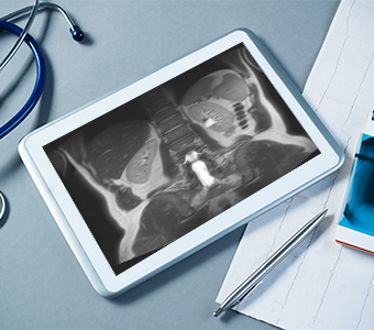
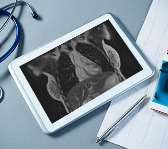
MRI of the pelvis
Do you need to prepare for the examination?
You must not eat for at least 6 hours before the examination. You may drink small amounts of water.
If you are having an examination of the rectum, you will need to use two laxative suppositories. Insert the first suppository 6 hours before the examination and the second 4 hours before the start of the procedure.
If you are booked for an examination of the cervix, gel will be applied into the vagina.
In all other respects, the general MRI guidelines apply.
How is the examination performed?
The radiographer will position you appropriately on the examination table and fit a coil for the part of the body being examined. You will also be given headphones to protect your hearing, as loud noises are heard during the examination. You will then be moved into the MRI machine for the duration of the scan. You will also be given a call bell, which you can press if you experience any problems during the procedure to contact the radiographer carrying it out.
It is especially important that you keep completely still during the examination itself, as movement degrades the quality of the images!
If there are significant motion artefacts from bowel movement due to active peristalsis, you will be given a medication to relax the bowel wall.
The examination takes about 45 minutes. If a contrast agent also needs to be administered, this adds a further 15 minutes.
Is a contrast agent required?
The decision to use a contrast agent depends on the indication for your examination and the findings during the scan itself. It is made by the radiologist supervising the examination.
MRI of the prostate
Do you need to prepare for the examination?
For the best results, it helps if your bladder is not completely empty, but you should not feel an urgent need to urinate or open your bowels. If you do, you should empty your bladder and bowels first.
In all other respects, the general MRI guidelines apply.
How is the examination performed?
The radiographer will position you appropriately on the examination table and fit a coil for the part of the body being examined. You will also be given headphones to protect your hearing, as loud noises are heard during the examination. You will then be moved into the MRI machine for the duration of the scan. You will also be given a call bell, which you can press if you experience any problems during the procedure to contact the radiographer carrying it out.
It is especially important that you keep completely still during the examination itself, as movement degrades the quality of the images!
If there are significant motion artefacts from bowel movement due to active peristalsis, you will be given a medication to relax the bowel wall.
The examination takes about 45 minutes.
Is a contrast agent required?
An intravenous contrast agent is always used for this type of examination.
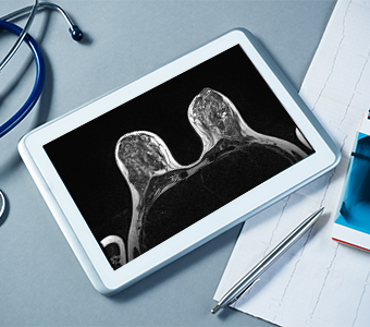
Breast MRI
When is the best time for a breast MRI?
If you have regular monthly periods, the ideal time for a breast MRI is between day 5 and day 15 of your menstrual cycle. The first day of bleeding counts as day 1.
If you are taking hormone replacement therapy, the contrast agent may accumulate in normal breast tissue, making the findings of the examination harder to interpret, so it is recommended that you stop the therapy at least 4 weeks before the examination.
An MRI is not advisable within 6 months of breast surgery or 12 months of breast radiotherapy, as changes related to healing can lead to a false positive result.
Do you need to prepare for the examination?
You must not eat for at least 6 hours before the examination.
In all other respects, the general MRI guidelines apply.
How is the examination performed?
Before the examination, we will insert a peripheral venous line for administering the contrast agent (usually in the crook of the elbow). The radiographer will position you on the examination table, lying on your stomach. Your breasts will be placed in the opening of the coil designed for breast examinations. You will be given headphones to protect your hearing, as loud noises are heard during the examination. You will also be given a call bell, which you can press if you experience any problems during the procedure to contact the radiographer carrying it out.
It is especially important that you keep completely still during the examination itself, as movement degrades the quality of the images!
The examination takes between 30 and 40 minutes.
Is a contrast agent required?
Yes, a breast MRI requires the use of an intravenous contrast agent.
MRI of the chest
Do you need to prepare for the examination?
If you are scheduled for an MRI of the chest with a contrast agent (for example, to characterise tumours, inflammation, etc.), do not eat for at least 6 hours before the examination.
In other cases, no special preparation is needed; only the general MRI guidelines apply.
How is the examination performed?
The radiographer will position you appropriately on the examination table (usually lying on your back) and fit a suitable coil. You will also be given headphones to protect your hearing, as loud noises are heard during the examination. You will then be moved into the MRI machine for the duration of the scan. You will also be given a call bell, which you can press if you experience any problems during the procedure to contact the radiographer carrying it out.
You will receive breathing instructions from the radiographer through the headphones (for example, “breathe in”, “breathe out” and “hold your breath”). In some scans, you may need to hold your breath for up to 15 seconds.
It is especially important that you keep completely still during the examination itself, as movement degrades the quality of the images!
The examination takes 20–30 minutes. If a contrast agent also needs to be administered, this adds a further 15 minutes.
Is a contrast agent required?
A contrast agent is used in these examinations only in certain cases, such as the characterisation of known tumours, inflammation, etc.
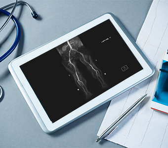
MR angiography (MRA)
What is MRA?
MRA is a diagnostic examination for imaging the blood vessels. It is used to detect vascular aneurysms and vascular malformations, to assess narrowing of vessels in atherosclerosis, to diagnose varicocele and pelvic congestion syndrome, etc.
Do you need to prepare for the examination?
If you have been referred for MRA of the abdominal vessels, do not eat for 6–8 hours before the examination.
No special preparation is needed for examinations of the blood vessels in other parts of the body; only the general MRI guidelines apply.
How is the examination performed?
The radiographer will position you appropriately on the examination table and fit a suitable coil for the part of the body being examined. You will also be given headphones to protect your hearing, as loud noises are heard during the examination. You will then be moved into the MRI machine for the duration of the scan. You will also be given a call bell, which you can press if you experience any problems during the procedure to contact the radiographer carrying it out.
It is especially important that you keep completely still during the examination itself, as movement degrades the quality of the images!
The examination takes around 45–60 minutes.
Is a contrast agent required?
A contrast agent is used in most of these examinations. However, the examination can also be performed without one, for example to assess vessel patency in the lower limbs in dialysis patients or young patients, to monitor the diameter of an abdominal aortic aneurysm, etc.

Appointments
We offer several ways to book an appointment. Choose the one that suits you best.
How to book
- In person – every working day during office hours: between 8:00 and 20:00.
- By post – to the address: MDT&T d.o.o. Lavričeva ulica 1, 2000 Maribor.
- Via our website or by email – at mr@mdt.si.
- By phone – every working day between 8:00 and 20:00 on 02 23 53 552 and 02 23 53 553.
You can also book healthcare services on the cakalnedobe.ezdrav.si and zvem.ezdrav.si websites.
Book an appointmentWaiting time
Waiting times for examinations for insured patients with a referral under the contract with ZZZS (the Health Insurance Institute of Slovenia), as at 2 June 2026 (the date is updated whenever a change occurs).
In accordance with the Rules on the maximum permissible waiting times for individual healthcare services and on the management of waiting lists, you can check waiting times on the website of NIJZ (the National Institute of Public Health).
Person responsible for the waiting list
Dr Marko Jevšek, MD, specialist radiologist
(mr@mdt.si, tel.: 02 23 53 552)
Tomaž Friedrich, MD, specialist radiologist
(mr@mdt.si, tel.: 02 23 53 552)
Waiting times
| Examination | Regular | Fast | Very fast |
|---|---|---|---|
| MRI of the head and neck | 90 days | 80 days | 70 days |
| MRI of the spine | 22 days | 20 days | 19 days |
| MRI of the knee; ankle; foot; shoulder; hand; wrist; elbow | 29 days | 27 days | 25 days |
| MRI of the hips; MRI of the skeleton without contrast agent – other | 50 days | 42 days | 35 days |
| MR arthrography of the hips | 62 days | 55 days | 36 days |
| MR arthrography of the shoulder | 34 days | 27 days | 20 days |
| MRI of the prostate | 126 days | 112 days | 105 days |
| MRI of the abdominal organs (upper abdomen; pelvis; urography; enterography) | 170 days | 160 days | 140 days |
| Breast MRI (depending on the patient's menstrual cycle) | 100 days | 80 days | 60 days |
| MR angiography | 85 days | 75 days | 60 days |
| MRI of the chest | 100 days | 80 days | 60 days |
Diagnostic imaging in the Diagnostični center Bled Group
The Diagnostični center Bled Group is the largest provider of diagnostic imaging in Slovenia. Every year we perform 130,000 MRI and 25,000 CT examinations on state-of-the-art equipment.
Appointments are available with a referral, for self-paying patients and for insurance companies.
Magnetic resonance imaging (MRI) is one of our main radiology services — within the Group it is performed at MDT&T and also at MTC Fontana in Maribor and Medilab in Ljubljana. At MDT&T we also perform X-ray examinations, while CT examinations (computed tomography) are performed at MTC Fontana and Medilab.
We have prepared summary tables to make booking MRI and CT examinations within the Group easier.
List of MRI examinations (PDF, in Slovenian) List of CT examinations (PDF, in Slovenian) Leaflet: MRI, CT and X-ray (PDF, in Slovenian)