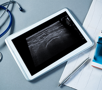
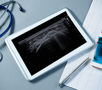
Ultrasound-guided procedures
Ultrasound-guided (US) procedures are minimally invasive procedures performed inside the body under ultrasound guidance. This allows us to puncture a joint, a calcification or diseased tissue with a fine needle with the greatest possible precision. They are performed under local anaesthesia and are used for both diagnostic and therapeutic purposes.
Ultrasound examinations are available on a self-pay basis only.
View the ultrasound price list Book an appointmentIs any preparation required before the examination?
No special preparation is required for ultrasound-guided procedures.
During the examination you will be asked to remove clothing from the area being examined.
What does the examination involve?
The doctor will position you appropriately on the examination table, clean the planned puncture site with a disinfectant and cover it with a sterile drape. Under ultrasound guidance, they will then puncture the site with a fine needle and carry out the appropriate procedure.
Pain during the procedure is usually minimal, as fine needles are used.
The treatment takes around 15–30 minutes.
If you have a blood-clotting disorder, diabetes or any kind of infection, you must let us know before your treatment.
Types of procedures
We perform the following ultrasound-guided procedures.
Ultrasound-guided (US) corticosteroid injection (block)
How is a US-guided corticosteroid injection performed?
The doctor first disinfects the puncture site and covers it with a sterile drape. They then insert a needle into the area where the corticosteroid is to be applied, using ultrasound guidance, which allows the needle tip to be positioned with maximum precision. The corticosteroid is then injected.
The procedure is performed under local anaesthesia.
Including preparation, it takes 5–10 minutes (the puncture itself usually less than 1 minute) and causes only minimal pain.
Are complications possible after the procedure?
The likelihood of complications is very low, and only mild forms are known.
A vasovagal reaction may occur during or immediately after the procedure.
Infection at the procedure site is also possible, but very rare.
What are the recommendations for patients after the procedure?
In the first two days after the treatment, shoulder pain may temporarily increase. This is a side effect of the corticosteroid. If this happens, we recommend taking an oral painkiller. The pain usually subsides on its own after some time.
In patients with diabetes, a corticosteroid injection may cause blood sugar fluctuations for up to a few days after the procedure. We therefore advise measuring your blood sugar more frequently.
If significant swelling, skin redness and pain develop, or if systemic signs of infection appear (such as a raised body temperature), seek medical attention as soon as possible.
If you have a blood-clotting disorder, diabetes or any kind of infection, you must let us know before your treatment.
Ultrasound-guided (US) lavage of calcifications
Calcific tendinosis is a condition characterised by the build-up of calcium in tendons, most commonly in the tendons of the shoulder muscles (the rotator cuff). Its cause is not yet understood. It mainly affects people of working age, most often women between the ages of 30 and 50. It may cause no clinical symptoms, but it frequently leads to pain and reduced mobility of the shoulder joint. In a certain proportion of patients, treatment with medication and physiotherapy is unsuccessful, so additional treatment is needed (US-guided lavage of the calcification, breaking up the calcification with shockwaves (ESWT) or surgical treatment), which in most cases significantly reduces or even eliminates the symptoms.
How is US-guided lavage of a calcification performed?
During the procedure, the doctor first punctures the calcification under US guidance. It is then flushed with saline solution in order to remove as much of it as possible. After this, an anti-inflammatory agent (a corticosteroid) is injected into the subacromial bursa to prevent subacromial bursitis, which can occur as a complication of the procedure itself. The aim of the treatment is to remove as much of the calcification as possible and thereby reduce its volume effect, which causes subacromial impingement and inflammation of the tendon in which the calcification is located. In this way, the clinical symptoms can be relieved to the greatest possible extent.
The procedure is performed under local anaesthesia.
Including preparation, it takes around 20–45 minutes.
Are complications possible after the procedure?
The likelihood of complications is very low, and only mild forms are known.
A vasovagal reaction may occur during or immediately after the treatment.
Within a few days or weeks after the procedure, subacromial bursitis may develop, presenting as a return of more pronounced pain in the shoulder area. In such cases, treatment with anti-inflammatory tablets (non-steroidal anti-inflammatory drugs) is required, or we may opt for a US-guided injection of an anti-inflammatory agent into the subacromial bursa.
Infection may occur at the procedure site, but this is very rare.
What are the recommendations for patients after the procedure?
In the first two days after the treatment, shoulder pain may temporarily increase. This is a side effect of the corticosteroid. If this happens, we recommend taking an oral painkiller. The pain usually subsides on its own after some time.
For 14 days, we advise against raising your arm above shoulder level or putting significant strain on the shoulder. After that, you can gradually start increasing your range of motion and the load on the shoulder.
In patients with diabetes, the corticosteroid injection may cause blood sugar fluctuations for up to a few days after the procedure. We therefore advise measuring your blood sugar more frequently.
If significant swelling, skin redness and pain develop, or if systemic signs of infection appear (such as a raised body temperature), seek medical attention as soon as possible.
If you have a blood-clotting disorder, diabetes or any kind of infection, you must let us know before your treatment.
Ultrasound-guided ganglion cyst aspiration
A ganglion cyst is a lump filled with viscous fluid. It is usually connected to a joint or tendon sheath via a thin stalk, which is why it most often lies next to joints or tendons. The wrist is a typical site; somewhat less commonly, it forms near the knee or ankle. It may cause pain that worsens with movement; less frequently, it presents as a painless lump. The cyst is not dangerous, but it can recur.
Indications for treating a ganglion cyst include pain, impaired function, signs of pressure on nerve structures and cosmetic reasons.
How is ultrasound-guided (US) ganglion cyst aspiration performed?
The doctor first disinfects the puncture site and covers it with a sterile drape. Using US guidance, they then puncture the ganglion cyst with a needle and drain it. If the cyst contents are very viscous, flushing may also be needed, and sometimes the cyst cannot be drained completely. A corticosteroid is then injected into what remains of the cyst.
The procedure is performed under local anaesthesia.
Including preparation, it takes 10–20 minutes and causes only minimal pain.
Are complications possible after the procedure?
The likelihood of complications is very low.
Infection or minor bleeding may occur at the wound site.
What are the recommendations for patients after the procedure?
In patients with diabetes, a corticosteroid injection may cause blood sugar fluctuations for up to a few days after the treatment. We therefore advise measuring your blood sugar more frequently.
If significant swelling, skin redness and pain develop, or if systemic signs of infection appear (such as a raised body temperature), seek medical attention as soon as possible.
If you have a blood-clotting disorder, diabetes or any kind of infection, you must let us know before your treatment.
Appointments
There are several ways to book an appointment for ultrasound-guided procedures. Ultrasound examinations are available on a self-pay basis only.
Radiology Clinic office hours: Mon–Fri 7:00–21:00 (bookings Mon–Fri 8:00–20:00).
- By phone: 02 23 53 552 or 02 23 53 553
- By email: mr@mdt.si
- In person at: Lavričeva ulica 1, 2000 Maribor
- By post to: MDT&T d.o.o., Lavričeva ulica 1, 2000 Maribor

Imaging diagnostics in the Diagnostični center Bled Group
In addition to basic diagnostics, doctors often recommend further imaging (MRI, CT or X-ray).
The Diagnostični center Bled Group is the largest provider of diagnostic imaging in Slovenia — appointments are available with a referral, for self-paying patients and for insurance companies.
Leaflet — MRI, CT and X-ray in the DCB Group (PDF, in Slovenian)